Cranial Cruciate Ligament Surgery
Surgery to treat lameness caused by Cranial Cruciate Ligament [CCL] trauma or disease is one of the most common orthopedic operations in dogs.
There are two cruciate ligaments within the stifle [knee] joint which act in opposite directions to each other to counteract shearing forces between the Femur [thigh bone] and Tibia [shin bone]. When damage occurs to these ligaments, it is almost always the cranial ligament that is affected (in purple below). The ligament can be torn by straining / twisting of the ligament during exertional exercise, but more commonly in the bigger breeds the ligament is gradually torn over a period of time – causing either continuous or intermittent lameness. It is now widely accepted that this is a ‘disease’ of the ligament rather than simple trauma. Unfortunately, once the ligament is damaged the gradual process of osteoarthritis [degenerative joint disease] will begin. Surgical treatment is designed to relieve as much pain and lameness as possible and reduce the onset and severity of arthritis.
Currently there are a number of surgical techniques used to treat these cases. They can be divided into techniques that Do or DON’T involve osteotomies [cutting into the bones].
- Without an osteotomy, Dr. Coudrai can offer the Lateral Suture Technique which, after inspection of the menisci [cartilages] in the joint and removal of any damaged sections, involves placing a monofilament nylon line from behind the lateral fabella to a small hole drilled in the tibial crest. The concept of this repair is to stabilize the joint with the prosthetic suture by placing it in a similar direction and taughtness to the original cruciate ligament. Eventually the prosthesis will break, but by then there should be enough fibrosis around the joint for it to function normally.
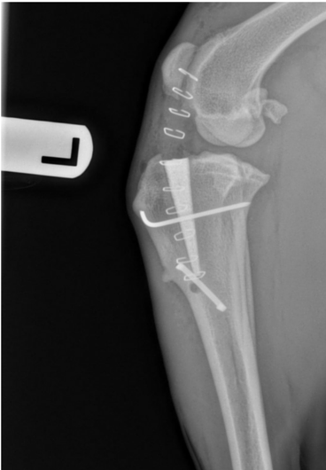
- Osteotomy techniques are now many and include tibial plateau leveling osteotomy [TPLO], tibial tuberosity advancement [TTA], triple tibial osteotomy [TTO], tibial wedge osteotomy [TWO] and others. These operations are all designed to remove the shear forces on the joint thus alleviating pain and lameness. They are all more complex procedures and inevitably involve the risk of bigger complications, but usually give a much better outcome and quicker return to function than lateral suture. Recently a variation of the TTA surgery has been developed called the Modified Maquet Procedure [MMP] using a Titanium Foam Wedge insert which is very exciting because the success rate is so high and the complication rate is much lower. Dr. Coudrai has performed over 100 surgeries with this technique and would recommend it as the best option for most cases. Comparison of MMP vs lateral suture technique results of surgeries performed by Dr. Coudrai has shown better lameness scores and weight bearing, as well as earlier return to function with significantly less post-operative pain with the MMP technique.


