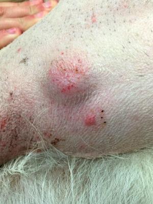
Mast Cells
Risk Factors for Mast Cell Tumors
Mast cell tumors occur commonly in dogs, less frequently in cats, and very rarely in human beings. They occur in dogs of all ages, breeds, and genders, anywhere in the body. Some breeds are predisposed to developing mast cell tumors, such as Beagles, Boston Terriers, Boxers, Bulldogs, Bullmastifs, Bull Terriers, Dachshunds, English Setters, Fox Terriers, Golden Retrievers, Labrador Retrievers, Schnauzers, American Staffordshire Terriers, and Weimaraners. Boxers are at the highest risk, though the tumors are often not as aggressive in this breed. Mast cell tumors may be associated with chronic immune over-stimulation that occurs in dogs with allergies or other inflammatory conditions. Older dogs are more likely to develop cancerous growths, with the average age of a dog with a mast cell tumor being 8-9 years old.
Symptoms
Symptoms can vary, depending on the location of the tumor and its degree of development. Signs may include:
- tumor
- loss of appetite
- vomiting
- bloody vomit
- diarrhea
- abdominal pain
- dark feces
- itchiness
- lethargy
- anorexia
- irregular hearth rhythm/blood pressure
- coughing
- labored breathing
- delayed wound healing
- bleeding disorders
- enlarged lymph nodes
Treating Mast Cell Tumors
The first step in treating a mast cell tumor is finding it. Hopefully the process can begin early when a Pet-Parent notices a growth on their dog. The veterinarian may use a fine needle to remove a sample for a preliminary biopsy prior to tumor removal. Blood tests may include a Complete Blood Count (CBC) and a serum chemistry profile. The CBC may show low or high white blood cell count, low platelet count, and elevated mast cell counts. Other tests may include urinalysis, x-rays, ultrasound, and aspirates of the lymph nodes or bone marrow.
The tumor is almost always surgically removed, if possible. The veterinarian performing the surgery removes a margin of healthy tissue around the tumor to capture any stray cancerous cells that may not be immediately obvious. After the tumor is removed, it is submitted for biopsy in a laboratory. One of the important aspects of the biopsy is determining whether or not the margins contain invasive cancerous cells.
The pathologist assigns a “grade” to the tumor, an assessment of how well differentiated the cells are and, by extension, how aggressively malignant the cancer appears to be. High-grade tumors may be treated with chemotherapy.

